Introduction to Low Vision
As a fully sighted individual, it can be difficult to understand why people with low vision see if very different ways to one another and to fully sighted people.
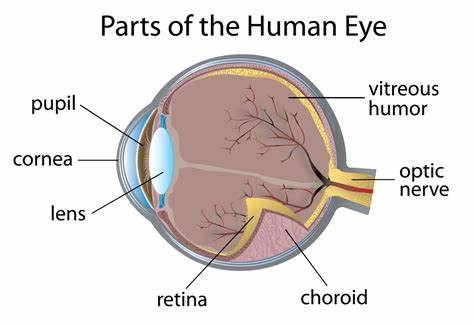
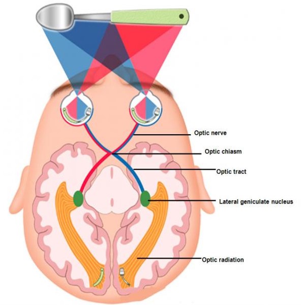
This unit will provide you with an overview of how the eye works, what can happen if parts of the eye aren't working properly and its impact on how people see in their day to day lives.
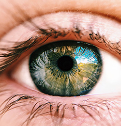
Overview of How the Eye Works
Click on the tabs to find different information
Possible Types of Low Vision
Click on the different headings to learn about possible types of low vision
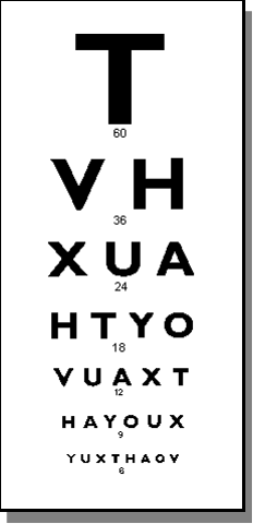
Visual acuity is our ability to see clearly and in detail. It is tested using a Snellen Chart.
The Snellen Chart measurement is used to determine a person's level of low vision.
The chart shows letters in decreasing size, with a number underneath each row of letters. (60, 36, 24, 18, 12, 9 and 6)
The testing distance from the chart is 6 meters. Results are displays as a fraction. The top number is the testing distance. The bottom number is how far down the chart the person is able to read.
Normal vision: 6/6 - This means that someone at a testing distance of 6 meters can read the bottom line on the chart (the line with 6 underneath).
Someone with a mild visual impairment would be able to read the fifth line down (line with a 12 underneath) so would have a reading of 6/12
Someone with a severe visual impairment would only be able to read the top line with the 60 underneath and so would have a reading of 6/60
If the top number could not been seen at 6 meter distance but at 3 meter distance, the reading would be 3/60, which is a profound visual impairement
Peripheral Vision Loss means that only central vision is retained.
Example illustrating a peripheral field loss:
Picture 1 is how a fully-sighted individual would see a picture of a dog.

Picture 2 is how someone with Peripheral Field Loss might see the same picture of the dog. This is known as tunnel vision.
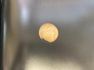
Remaining Vision: Tunnel Vision ("What" Vision)
- Photoreceptor cones are working but there are problems with the rods
- Cones need light to work and are responsible for colour and detail
- With Tunnel Vision, someone may be able to read, do detailed tasks and see colour
- However, someone will experience night blindness, difficulty adjusting to changes in lighting levels and in bright sunshine
- Movement can be difficult as obstacles disappear or suddenly appear
- Also, people can appear without warning if they approach from the person from the side
- Standing to close to the person will mean than they may not be able to see your entire face which makes communication difficult (Too close; too big)
Central Field Loss means that only peripheral vision is retained.
Example illustrating a central field loss:Picture 1 is how a fully-sighted individual would see a picture of two children holding balls.
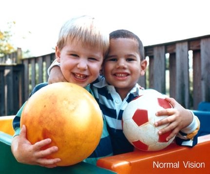
Picture 2 is how someone with Central Field Loss might see the same picture the two boys.
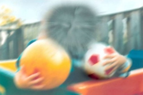
Remaining Vision: Peripheral Vision ("Where" Vision)
- Photoreceptor rods are working but there are problems with the cones
- Rods work in low light and are responsible for shape, shadow and movement
- The central loss may appear like an image vacuum/absence or as a scotoma which appears like a dark blob
- Moving might be OK as someone has enough vision to see and avoid obstacles and possibly detect steps but will experience difficulty with detailed tasks such as reading, identifying colours and recognising faces
- The person may adapt unusual head positions and task position to make best use of their vision, this is called eccentric fixation
- The person may not make eye contact in attempt to see faces
Central and Peripheral Field Loss is complex and results from damage to different parts of the eye impacting both visual fields.
- Cause of loss may be situated at the front or back of the eye or along the visual pathway
- May have any one of or combination of types of vision depending on cause of loss – Distorted vision, blurred vision, patchy vision, altered colour vision, half/quarter visual field, light perception, total loss of sight

Let's Review!
Click on the correct option
What type of vision does this image depict?

Click on the correct option
What type of vision does this image depict?

Click on the correct option
What type of vision does this image depict?


